Epsilon Dermalyser New features include:
a) adjustable rectangular RoI (Region of Interest)
b) image builder tool for larger area skin images
c) 3-D image presentation that emphasises skin surface texture
Rapid Matching
Rapid matching of two or more images of the same skin area forms a powerful analysis tool. The user selects any representative region in an image, then the software searches and locates identical regions in subsequent images. This is shown in Figure 1 where, even though hairs (thick dark lines) are displaced between images, the software finds the same RoI position relative to skin features (thin lines) in the other three images.
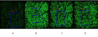
Figure 1. Matching of Epsilon skin images. A Region of Interest is selected in image B and the same region is identified in images A, C and D. Image A is pre-treatment and B, C and D are post-treatment.
This quick and simple skin matching capability enables more accurate and productive analysis of skin image dynamics over hours, weeks or years. This is crucial for such topics as skin wound healing, skin disease treatment, and trans-dermal drug delivery.
Large area skin images
Large area skin images are built by stitching a linear image sequence together. The user selects a region within an initial image and the Epsilon Dermalyser Image Analysis software locates the identical overlapping region belonging to the image of an adjacent skin location. Using the initial RoI, the two images are automatically combined along one edge. This can be repeated for any sequence of images, joining either top, bottom, left or right edges, as depicted in Figure 2.

Figure 2. The original Epsilon skin images (top), and the stitched Epsilon skin image (bottom).
The top sequence in Figure 2 shows the original discrete Epsilon skin images. Beneath that are the same images combined across a large area of skin. Such images can be highly valuable for cosmetic and pharmaceutical studies.
Apart from the features presented above, Epsilon Demalyser™ can also render images in 3D for enhanced visualisation of skin surface texture.
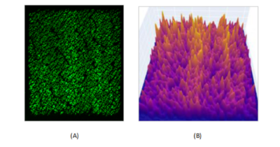
Figure 3. Epsilon’s skin capacitance 2D image (A) and the Dermalyser 3D display (B).
Figure 3 above shows a skin site with two ambiguous vertically oriented regions of increased hydration, and its corresponding 3D-enhanced display.